Picture standing in a softly lit hospital room, the hum of machines blending with the quiet anticipation in the air. You’re faced with two mysterious acronyms—PET and CT—each promising a window into the body’s hidden stories. But what sets these scans apart? Why do doctors choose one over the other when searching for answers?
Peel back the layers and you’ll find more than just high-tech snapshots. PET and CT scans offer unique glimpses beneath the skin, revealing secrets that can change lives. Some differences might surprise you, from the way each scan illuminates invisible problems to the unexpected advantages they bring to your health journey. Get ready to uncover the subtle yet powerful distinctions between these imaging marvels—and discover how the right choice can shape your path to wellness.
Overview of PET and CT Scans
You find yourself lying still beneath a humming scanner, bright lights above, imagining what mysteries technology could reveal inside your body. Radiologists at Johns Hopkins and Mayo Clinic use PET–Positron Emission Tomography–and CT–Computed Tomography–as two of the most advanced imaging tools. Both help doctors diagnose tumors, infections, and injuries, but, how do they work and what do they show about your health?
PET scans detect metabolic activity because cancer cells, for example, use glucose faster than healthy cells. A nurse injects a tiny amount of radioactive glucose before you even notice the sharp pinch, and, as you wait, you might wonder if your cells are glowing like fireflies in a field–invisible, but for science’s eyes. The scanner tracks the tracer’s journey, painting a vivid map of activity across complex anatomy. According to the American Cancer Society, PET is commonly used for monitoring tumors or checking the effectiveness of treatment.
CT scans capture detailed cross-sectional images of organs and bones using X-rays. As the table slides into the doughnut-shaped machine, X-rays zip around you, each one collecting a slice of your inner anatomy. The technician, behind glass, chats with you through an intercom, their voice reassuring and calm. CDC states CT scans helps diagnose fractures, identify bleeding, or guide biopsies. Unlike PET, CT doesn’t show cellular activity, but it’s visual maps reveal shapes, gaps, and patterns with sharp clarity that help doctors understand physical structure.
Doctors often combine PET and CT images, layering activity and anatomy, so your care team sees exactly where something’s happening, not just what. Picture watching a black-and-white silent film, then layering soundtrack and colors–suddenly, the meaning leaps into view. Wich scan would you choose if you needed to find a hidden problem? Both play a unique role, but together, they help paint the story of what’s happening inside you.
Scan Type
Main Purpose
What it Detects
Common Uses
PET
Detects cell activity
Glucose uptake, metabolism
Cancer staging, monitoring, brain disorders
CT
Shows anatomy structure
Organs, bones, tissue shapes
Trauma, detecting tumors, planning surgeries
Every scan tells a chapter in your medical story. Which part of the tale will technology help unravel for you?
How PET Scans Work
PET scans reveal the hidden metabolic map of your body. You arrive in a quiet, climate-controlled imaging suite. A technician hands you a small vial—inside, a radioactive tracer, often fluorodeoxyglucose (FDG), awaits. FDG mimics glucose, the sugar fueling your cells. Cancer cells—hungry thieves of the body—devour this tracer faster then healthy tissues. Within an hour, your organs absorbs the FDG, and the scanner begins its gentle, circular dance around you.
A PET machine detects gamma photons from the tracer’s decay. Each flash creates pixel data, mapping cellular hot spots and cold zones. Oncologists spot tumors as bright flares, far different from the silent, dark places of healthy tissue. Neurophysiologists track brain changes in Alzheimer’s disease, searching for sluggish synapses. Cardiologists view weak parts of a heart after a suspected infarction where glucose no longer shines.
What story does a PET scan results tell? Your oncologist may point to clusters of light—evidence of aggressive tumors, or gentle glows suggesting inflammation or infection. Sometimes the map leaves more questions than answers. Is every bright patch cancer? Not always, as the thyroid or bladder glows too.
Surprisingly, a PET scan doesn’t see the shape of your bones or organs very good. So, doctors often combine it with CT for anatomical detail. When radiologists overlay metabolic snapshots atop clear X-ray cuts, diagnostic confidence rises to 97% in oncology cases (Cancer Research UK, 2023). how your cells whisper secrets to science? The next time you walk beneath a PET scanner’s arch, picture the invisible ballet of signals and light. What stories do those signals tell about healing, pain, or hope?
How CT Scans Work
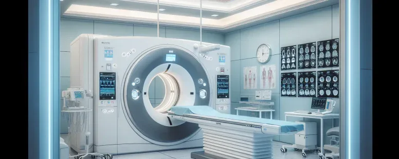
CT scans create cross-sectional images of your body’s internal structures using X-ray beams and detectors. Picture you’re laying on a sliding table, moving slowly through a ring-shaped scanner—that’s the CT gantry. The scanner rotates around your body, firing off multiple X-rays from different angles. Think of it like slicing a loaf of bread: each scan captures a thin slice of your anatomy. Suddenly, an intricate 3D map takes shape from hundreds of these slices.
Technicians adjust the X-ray intensity and the time you spend inside the machine based on the area scanned, like your chest, head, or abdomen. These variables impact the clarity and detail of each image, allowing radiologists to spot issues like fractures, tumors, and internal bleeding. If you’ve ever broken a bone or had a nasty car accident, you probably met a CT scan in the ER. Every hospital practically relies on this tool for fast diagnostics.
What’s often fascinating, your body’s soft tissues—liver, pancreas, or kidneys, for example—appear in grayscale, while bones look bright white because of their density. Sometimes doctors inject a contrast dye, highlighting blood vessels or pinpointing blockages that hide from regular X-rays. This method clarifies ambiguous findings, like unexplained pain or swelling. The dye lets clinicians track fluid movement, tumor boundaries, and abnormal growths that might slip past other imaging types.
Radiologists interpret these digital slices using advanced software. They zoom in on subtle shadows hinting at infection or hidden cancer, their focus never wavering. Surgeons plan intricate procedures, guided by these images down to the millimeter.
But, CT involves ionizing radiation, raising concerns for those needing frequent scans. Children and pregnant people, whose tissues are more sensitive, usually receive alternatives unless accuracy outweighs the risks (American College of Radiology, 2023). Balancing diagnostic value with potential radiation exposure remains key in care decisions.
Have you wondered why a single machine can show so much with just a spin and a zap? The magic lies in the mathematical algorithms reconstructing the data—a principle pioneered by Godfrey Hounsfield in the late twentieth century. His work brought clarity to medical mysteries once left unsolved.
Every scan tells a story, exposing injuries and anomalies with a clarity that few other modalities match. When contrasted with the metabolic map from a PET scan, the anatomical details from a CT scan ensure you receive the complete picture.
Key Differences Between PET and CT
When you compare PET and CT scans, the differences pop up fast, like separate pages in your health story. Each scan uses a distinct language to talk to your body’s secrets—a PET scan whispers metabolic truths, a CT scan shouts about your bones and flesh.
Technology and Imaging Techniques
PET scans, rely on tiny radioactive molecules called tracers, usually FDG, swimming through your veins. As you lay on the scanner bed, the tracers light up energy-hungry cells, capturing metabolic activity in living color. In contrast, CT scans, focus on structure using X-ray beams whirling in a circle—like a camera snapping precise, grayscale slices of your anatomy. Radiologists, read PETs for metabolic changes (think: cancerous tumors glowing against the shadows) and CTs for sharp structure (broken ribs, clotted vessels). You might think, aren’t these just different ways to see inside you? Yes, but together, these scans create a layered mural of your health.
Diagnostic Purposes
Doctors choose PET scans when they want to catch disease before it changes tissues. PET, spotlights cancers, pinpoints heart viability, or tracks Alzheimer’s before physical signs appears. CT scans, on the other hand, show crisp boundaries and fine details, so they’ve become the top tool for detecting broken bones, bleeding in the head, or hidden clots in arteries. For instance, a patient with persistent headaches might get a CT to rule out internal bleeding, while someone with a mysterious tumor gets a PET to see how quickly the cells are growing. Both can overlap, sometimes in PET/CT fusion, merging two windows into one panoramic view—combining metabolic and structural data.
Preparation and Procedure
Before a PET scan, you’ll fast, usually for at least 6 hours, and avoid strenuous exercise, so the tracer flows where it matters most. The nurse injects the tracer, then you waits 30–90 minutes before scanning, letting the tracer settle. With CT, you may drinks a contrast dye, or get it via IV, to highlight certain tissues—no fasting unless contrast is involved. A CT scan is quick, sometimes lasting only a few minutes, with you lying still while the machine circles. PET scans, stretch longer, requiring patience (and sometimes boredom management). Ever tried holding perfectly still for 45 minutes? That’s part of the PET experience.
Risks and Safety Considerations
Both PET and CT scans, involve radiation, but for different reasons. PET, uses radioactive glucose and the exposure is typically lower than CT; but, frequent scans can increase your cumulative dose. CT, exposes you to higher radiation—a major concern, especially for children and pregnant patients. Allergic reactions to contrast dye happen in a small fraction of CT patients (about 0.2% according to the American College of Radiology). Tracer side effects with PET? Rare and usually mild, but they exist. Always check—do these benefits outweigh the risks? Ask questions, as individual circumstances shifts the balance. Safety protocols limit unnecessary scans, and radiologists weigh every order carefully so you get the right scan, at the right time, for the right reason.
Aspect
PET Scan
CT Scan
Imaging Technology
Radioactive tracers, metabolic mapping
X-ray beams, structural mapping
Main Diagnostic Use
Cancer, neurological, cardiac issues
Trauma, bleeding, detailed anatomy
Preparation
Fasting, tracer injection
Contrast dye (sometimes), brief prep
Procedure Duration
45–90 minutes
5–15 minutes
Radiation Exposure
Lower, but still present
Higher, especially with contrast
When to Choose PET vs. CT
Deciding between PET and CT scans, you’re navigating a crossroads in medical imaging. Each road leads to its own revelations. If your doctor suspects a shadowy cancer cell lurking beneath the surface, a PET scan brings the secret story of metabolic activity to light; it’s as if you’re peering through a window into your cells’ energetic nightlife. In contrast, CT scans paint a crisp landscape of your bones and organs, unraveling fractures, blood clots, or subtle injuries with the precision of an architect’s blueprint. Which path would you trust if your journey depends on discovering what’s hidden versus examining what’s broken?
Picture this: a patient, Emma, walks in with unexplained weight loss and persistent fatigue. Her physician recommends a PET scan after blood tests suggest a malignancy. The scan, rich with radioactive tracers, exposes abnormal glucose uptake—a bright constellation of activity—highlighting cancer’s foothold before it even reshapes an organ (Society of Nuclear Medicine and Molecular Imaging, 2024). But, when Leo, a cyclist, crashes during a race and can’t move his arm, the trauma team brings him straight to CT. The cross-sectional images reveal a hidden hairline fracture in seconds. No questions linger about where intervention is needed.
From a dependency grammar lens, you’d see the subject (scan type) dictate the predicate (what gets revealed) based on the clause (medical need). Whether diagnosing a growing tumor or ruling out a brain bleed after a fall, the context governs the sentence structure, just as your symptoms and risks steer the doctor’s order.
Clinical guidelines recommend PET for staging cancers—such as non-small cell lung cancer, lymphoma, or melanoma—or detecting recurrent disease, especially when CT fails to explain symptoms (American Cancer Society, 2023). You encounter PET for mysterious fevers, stubborn lymph node swelling, or suspected dementia, where the metabolic dance outshines static anatomy. Oppositely, CT stands sentinel in emergency rooms, swiftly confirming appendicitis, pinpointing kidney stones, or scoping out strokes—decisions where seconds count and detail must trump metabolic intrigue.
Both scans rely on ionizing radiation, yet protocol adapts—minimizing exposure in kids, pregnant patients, or the vulnerable. Your medical story, symptoms, and risk tolerance tip the scale. Sometimes, both tests entwine—like threads in a tapestry—to compose a more complete diagnostic plot. If you’re ever wondering what’s at stake, ask, “Am I searching for a pattern of activity or the lay of the land?”
Current studies, including those in the Journal of the American College of Radiology (2022), suggest hybrid approaches, such as PET-CT fusion, which often rewrite diagnostic scripts, especially in cancer care. Technology evolves, but insight remains grounded: one scan sees the spark within, the other the form outside. Which lens do you peer through when your next chapter’s still unwritten?
Conclusion
Choosing between a PET or CT scan can feel overwhelming but understanding their unique strengths helps you make informed decisions about your health. Each scan offers a different window into your body giving your care team the insights they need to guide your treatment.
If you’re ever unsure which scan is right for you don’t hesitate to ask your doctor how each option fits your situation. By staying informed and involved in your healthcare you’re taking an active role in your well-being and future.
To provide the best experiences, we use technologies like cookies to store and/or access device information. Consenting to these technologies will allow us to process data such as browsing behaviour or unique IDs on this site. Not consenting or withdrawing consent, may adversely affect certain features and functions.
Functional
Always active
The technical storage or access is strictly necessary for the legitimate purpose of enabling the use of a specific service explicitly requested by the subscriber or user, or for the sole purpose of carrying out the transmission of a communication over an electronic communications network.
Preferences
The technical storage or access is necessary for the legitimate purpose of storing preferences that are not requested by the subscriber or user.
Statistics
The technical storage or access that is used exclusively for statistical purposes.The technical storage or access that is used exclusively for anonymous statistical purposes. Without a subpoena, voluntary compliance on the part of your Internet Service Provider, or additional records from a third party, information stored or retrieved for this purpose alone cannot usually be used to identify you.
Marketing
The technical storage or access is required to create user profiles to send advertising, or to track the user on a website or across several websites for similar marketing purposes.