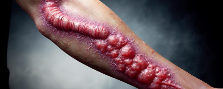
When you notice an unusual raised scar forming after an injury, it might be more than just normal healing. Keloid and hypertrophic scars, while similar in appearance, represent two distinct responses to skin trauma. Understanding the difference between these two types of scars isn’t just about terminology—it’s crucial for proper treatment and management.
While both conditions involve excess collagen production during the healing process, they behave differently over time. Keloids aggressively extend beyond the original wound boundaries and rarely improve without intervention, while hypertrophic scars remain within the injury’s borders and may improve naturally over months or years. These differences in behavior, appearance, and treatment options make accurate identification essential for anyone dealing with problematic scarring.
What Are Scars and Why Do They Form?
Scars develop as part of your body’s natural healing process after skin damage. They’re composed of fibrous tissue that replaces normal skin following an injury, surgery, or disease. When your skin suffers damage, your body immediately begins a repair process that involves several phases.
The healing process starts with inflammation, where blood clotting forms a protective scab. Next, fibroblast cells produce collagen to rebuild the damaged area. Finally, the remodeling phase occurs where collagen fibers reorganize and strengthen the new tissue.
Scar formation is influenced by several factors:
Wound depth (deeper wounds typically create more prominent scars)
Location on the body (areas with high tension or movement scar more visibly)
Age (younger skin tends to heal differently than older skin)
Genetics (your family history affects how you scar)
Skin type and ethnicity (darker skin types are more prone to certain scar types)
Your body produces collagen to repair wounds, but sometimes this process doesn’t work perfectly. When collagen production continues beyond what’s needed or occurs in a disorganized pattern, problematic scars like keloids or hypertrophic scars can form.
The normal scar formation process completes in approximately 12 to 18 months, at which point most scars fade and flatten naturally. But, in some cases, the healing process goes awry, resulting in abnormal scarring that’s more noticeble and sometimes uncomfortable.
Understanding why scars form is crucial for addressing different scar types and determining appropriate treatment approaches. While all scars result from the same basic healing mechanism, the variations in this process lead to the different types of scars you might experience after an injury.
Understanding Hypertrophic Scars
Hypertrophic scars are raised, reddish marks that form within the boundaries of the original wound. Unlike keloids, these scars don’t extend beyond the injury site and often improve gradually over time without intervention. Hypertrophic scarring occurs in approximately 40-70% of surgical incisions and up to 91% of burn injuries, making them more common than keloids.
Characteristics of Hypertrophic Scars
Hypertrophic scars have distinct features that differentiate them from other scar types. They’re typically red or pink, raised above surrounding skin, and remain confined to the original injury borders. These scars feel firm and may appear thickened due to excessive collagen deposition. Hypertrophic scars often develop within 4-8 weeks after wound closure and reach their maximum size within 6 months. They’re more likely to form over joints or areas with high tension and movement, such as the shoulders, neck, or knees. Unlike keloids, hypertrophic scars frequently show spontaneous regression after 6-12 months, gradually flattening and fading though they rarely disappear completely.
Common Causes and Risk Factors
Several factors increase your likelihood of developing hypertrophic scars. Wounds with delayed healing (taking longer than 3 weeks to close) create a 15-20% higher risk of hypertrophic scarring. Burns represent the most common cause, with studies showing 30-90% of burn injuries developing these raised scars. Surgical incisions, especially those placed perpendicular to natural skin tension lines, have increased susceptibility to hypertrophic formation.
Your personal risk factors include age (younger individuals heal with more robust collagen production), genetic predisposition, skin type (darker skin tones have 15-20% higher incidence), and hormonal influences. Infections or wounds under tension significantly increase hypertrophic development risk. People with compromised immune systems also experience different healing responses that can lead to abnormal scarring.
Environmental factors play a role too. Wound location matters significantly—areas with high skin tension or frequent movement, such as the chest, shoulders, and back, experience hypertrophic scarring at rates 3-5 times higher than other body regions. Early and appropriate wound care reduces your risk by minimizing inflammation and supporting optimal healing conditions.
Understanding Keloid Scars
Keloid scars represent a unique type of abnormal scarring characterized by excessive growth beyond the original wound boundaries. Unlike typical scars, keloids continue to expand over time and rarely regress naturally, making them particularly challenging to treat effectively.
Characteristics of Keloid Scars
Keloid scars exhibit distinctive features that set them apart from other scar types. These raised, firm growths extend beyond the original wound borders, appearing smooth, shiny, and dome-shaped with a rubbery texture. Keloids typically appear pink to purple or darker than surrounding skin, depending on your skin tone. They’re often accompanied by symptoms like tenderness, itchiness, or pain when touched, and may cause a pulling sensation as they continue growing. Unlike hypertrophic scars, keloids don’t shrink over time and can develop months or even years after the initial injury has healed.
Common Causes and Risk Factors
Keloids form from an abnormal healing response where fibroblast cells produce excessive collagen after skin injury. Common triggers include surgeries, ear piercings, acne, burns, chickenpox, and even minor scratches or insect bites. Your risk of developing keloids increases significantly with certain factors. People with darker skin tones, particularly those of African, Asian, or Hispanic descent, face 15-20 times higher keloid risk. Genetic predisposition plays a crucial role – keloids runs in families with approximately 50% of affected individuals reporting a family history. Age also matters, with individuals between 10-30 years experiencing peak vulnerability. Specific body areas such as the chest, shoulders, earlobes, and upper back are more susceptible to keloid formation due to high skin tension in these regions.
Hormone fluctuations during puberty or pregnancy can exasperate keloid development, which explains why some women notice keloid formation or growth during these periods. Environmental factors like repeated trauma to the same area or wound infections that prolong the inflammatory phase also increase your likelihood of developing these problematic scars.
Key Differences Between Keloid and Hypertrophic Scars
Keloid and hypertrophic scars differ in several critical ways that affect their management and treatment outcomes. Understanding these distinctions helps healthcare providers determine appropriate treatment approaches and helps patients recognize what type of scar they’re dealing with.
Appearance and Growth Patterns
Keloid scars exhibit distinctive growth patterns that separate them from hypertrophic scars. Keloids appear as smooth, hard, raised growths that extend beyond the original wound boundaries, often resembling a claw or tentacle-like formation. They typically start as small, firm nodules that progressively enlarge over months or years, sometimes reaching several centimeters in size. Their color ranges from pink to purple or dark brown, depending on the patient’s skin tone.
Hypertrophic scars present as raised, red, and firm tissue that remains confined within the original wound margins. Unlike keloids, these scars don’t invade surrounding healthy tissue. Hypertrophic scars generally reach their maximum size within 6 months and begin to regress afterward, often flattening and fading significantly by 12-18 months without intervention. Their appearance is typically more regular and follows the shape of the original injury.
Location and Distribution
Keloids and hypertrophic scars tend to develop in different body areas with varying frequency. Keloids commonly form on the earlobes, shoulders, chest, back, and jawline—areas with high skin tension or frequent movement. These locations experience consistent stress during daily activities, potentially contributing to abnormal collagen production. Some patients develop keloids exclusively in certain body regions while healing normally elsewhere.
Hypertrophic scars can occur anywhere on the body but are most prevalent in areas crossing joint spaces or regions subject to tension during wound healing. Common sites include the shoulders, neck, knees, and elbows. They’re particularly common in burn injuries across flexor surfaces where skin naturally stretches and contracts. Unlike keloids, hypertrophic scars don’t show the same predilection for specific anatomical regions and distribute more according to injury patterns rather than genetic tendencies.
Genetic Factors
Genetic predisposition plays a significant role in determining scar type formation. Keloid development has a strong genetic component, with 50-80% of patients reporting a family history of keloid formation. Studies have identified several genetic markers associated with keloid formation, including variations in the HLA-DRB1, NEDD4, and FOXL2 genes. Certain ethnic groups demonstrate higher susceptability to keloid formation, particularly individuals of African, Asian, and Hispanic descent, who are 15-20 times more likely to develop keloids than those with lighter skin tones.
Hypertrophic scarring shows less pronounced genetic correlation compared to keloids. While some familial tendencies exist, the genetic links aren’t as clearly defined. Environmental factors and wound management tend to play a more dominant role in hypertrophic scar development than genetic predisposition alone. The difference in genetic influence explains why some patients consistently form keloids following any skin injury, while hypertrophic scarring typically depends more on the nature and management of specific wounds.
Diagnosis and Medical Assessment
Differential Diagnosis Techniques
Medical professionals use specific diagnostic techniques to differentiate between keloid and hypertrophic scars. Visual examination serves as the primary assessment method, with doctors evaluating the scar’s boundaries relative to the original wound. Keloids extend beyond wound borders, while hypertrophic scars remain within them. Dermatologists often document the scar’s color, texture, and elevation to track changes over time.
Dermoscopy enhances diagnostic accuracy by magnifying surface details of the scar tissue. This non-invasive technique reveals vascular patterns specific to each scar type – keloids typically display fewer blood vessels with irregular distribution, whereas hypertrophic scars show more organized vascular structures. The duration and progression of the scar provide additional diagnostic clues, as hypertrophic scars usually develop earlier (4-8 weeks post-injury) compared to keloids (months or years after injury).
Biopsy and Histological Examination
A skin biopsy offers definitive differentiation between keloid and hypertrophic scars when visual assessment proves inconclusive. During this procedure, doctors remove a small sample of scar tissue for laboratory analysis. Histological examination reveals distinctive cellular and structural patterns in the tissue samples.
Under microscopic examination, keloids contain thick, haphazardly arranged collagen bundles that extend into surrounding normal tissue. These collagen fibers appear as whorls or nodules with excessive mucin and glycosaminoglycans between them. Hypertrophic scars, by contrast, show more parallel collagen fiber alignment confined to the dermis layer of the injury site. The presence of myofibroblasts (specialized contractile cells) differs between the two scar types – they’re abundant in early hypertrophic scars but less prominent in mature keloids.
Medical Imaging and Assessment Tools
Advanced imaging techniques support accurate diagnosis of challenging scar cases. High-frequency ultrasound measures scar thickness and density, revealing deeper structural differences between keloid and hypertrophic tissue. Keloids typically appear more heterogeneous on ultrasound with greater thickness measurements.
Several validated assessment scales help quantify scar characteristics and monitor treatment progress. The Vancouver Scar Scale (VSS) evaluates vascularity, pigmentation, pliability, and height of scars on a numerical scale. The Patient and Observer Scar Assessment Scale (POSAS) incorporates both clinician observations and patient-reported symptoms such as pain, itching, and functional limitations. These standardized tools ensure consistent documentation across different healthcare providers and treatment sessions.
When to Seek Medical Evaluation
You should consult a dermatologist when your scar exhibits concerning changes such as rapid growth, persistent pain, or restricted movement. Early assessment improves treatment outcomes, particularly for individuals with risk factors for abnormal scarring. Dermatologists often recommend preventive strategies for patients with a history of keloids before undergoing procedures like piercings or elective surgeries.
A comprehensive medical evaluation includes review of your medical history, family history of abnormal scarring, and detailed documentation of the original injury. Doctors evaluate functional impairment caused by the scar, such as limited joint mobility or sensory changes. The psychological impact of visible scarring also factors into treatment planning, as both keloid and hypertrophic scars can cause significant emotional distress and affect quality of life.
Treatment Options for Hypertrophic Scars
Hypertrophic scars respond well to various treatment methods, with options ranging from non-invasive approaches to surgical interventions. Effective treatment plans typically combine multiple modalities tailored to the scar’s characteristics, location, and the patient’s specific needs.
Topical Treatments
Silicone-based products form the cornerstone of hypertrophic scar management. Silicone gel sheets and silicone gels create a semi-occlusive barrier that hydrates the scar tissue and reduces collagen production. Clinical studies show 60-85% improvement in scar thickness and redness when silicone sheets are applied 12-24 hours daily for at least 2-3 months. Other topical options include:
Pressure garments that flatten scars by applying 20-30 mmHg of pressure
Corticosteroid creams that reduce inflammation and inhibit collagen synthesis
Onion extract gels containing antioxidants and anti-inflammatory compounds
Vitamin E preparations that may improve scar appearance through moisturization
Injectable Treatments
Intralesional corticosteroid injections effectively reduce hypertrophic scar elevation and symptoms. Triamcinolone acetonide (10-40 mg/ml) injected directly into the scar at 4-6 week intervals shows significant improvement in 50-100% of cases. Common injectables include:
5-Fluorouracil (5-FU) that inhibits fibroblast proliferation
Bleomycin that degrades collagen fibers and induces apoptosis
Botulinum toxin that reduces muscle tension around healing wounds
Platelet-rich plasma containing growth factors that promote proper healing
Laser and Light Therapies
Laser treatments target the vascular and collagen components of hypertrophic scars. Pulsed dye lasers (PDL) at 585-595 nm wavelengths reduce redness by targeting hemoglobin in blood vessels. Fractional lasers create microscopic treatment zones that stimulate collagen remodeling. Light-based options include:
CO₂ lasers that vaporize scar tissue and promote reorganization
Nd:YAG lasers that penetrate deeper into the dermis for collagen remodeling
Intense pulsed light (IPL) systems that reduce redness and improve texture
LED phototherapy that modulates cellular activity and reduces inflammation
Surgical Approaches
Surgical intervention is considered for hypertrophic scars resistant to other treatments. Techniques such as scar revision involve excising the scar and reclosing the wound using advanced suturing methods. Timing is crucial, with procedures ideally performed after the scar has matured (typically 12-18 months post-injury). Surgical options include:
Z-plasty that changes the scar direction to aligns with natural skin lines
W-plasty that breaks up linear scars into irregular patterns
Tissue expansion that stretches normal skin for reconstruction
Skin grafting for covering large areas after scar excision
Combination Therapy
Most hypertrophic scars benefit from multimodal approaches. Combining silicone sheets with pressure therapy increases effectiveness by 90% compared to either treatment alone. Similarly, using corticosteroid injections before laser therapy reduces treatment sessions by 30-50%. An experienced dermatologist or plastic surgeon can develop a customized treatment plan based on the scar’s characteristics and patient goals.
Treatment Options for Keloid Scars
Medical Treatments
Keloid scars require specialized medical interventions due to their persistent growth patterns. Intralesional corticosteroid injections represent the first-line treatment, reducing inflammation and inhibiting collagen synthesis in 70-100% of cases. These injections are typically administered every 4-6 weeks until the keloid flattens. Cryotherapy freezes and destroys keloid tissue, providing up to 73% improvement when combined with steroid injections. For extensive keloids, 5-Fluorouracil injections target fibroblast proliferation, while interferon injections inhibit collagen production in treatment-resistant cases.
Surgical Approaches
Surgical excision of keloids must be approached cautiously as 45-100% of keloids recur after surgery alone. Modern techniques combine excision with adjunctive treatments like post-operative radiation therapy, which reduces recurrence rates to 10-20%. Laser therapy, particularly pulsed-dye laser and CO2 laser treatments, effectively reduces keloid redness, thickness, and associated symptoms. These surgical interventions work best when incorporated into comprehensive treatment plans rather than as standalone procedures.
Non-Invasive Options
Several non-invasive treatments provide relief without surgical intervention. Silicone gel sheets and gels create a protective microenvironment that hydrates and softens keloids when applied for 12-24 hours daily over 2-3 months. Pressure therapy using custom-made compression garments exerts 24-30 mmHg of pressure on keloids, helping to reorganize collagen fibers and flatten the scar. These approaches work best for smaller keloids or as maintenance therapy following more aggressive treatments.
Emerging Therapies
Innovative approaches for keloid management include botulinum toxin (Botox) injections, which reduce tension around wounds and inhibit fibroblast proliferation. Bleomycin, an antitumor agent, decreases keloid volume by up to 92% after multiple treatments. Imiquimod cream stimulates the immune system to break down abnormal scar tissue, while tacrolimus ointment targets the inflammatory pathways involved in keloid formation. These emerging options offer hope for patients with recalcitrant keloids that haven’t responded to traditional treatments.
Combination Therapy
The most effective keloid treatment protocols use combination approaches tailored to individual scar characteristics. A typical regimen might include surgical excision followed by immediate corticosteroid injection, silicone sheeting, and pressure therapy. This multi-modal strategy addresses different aspects of keloid formation, including inflammation, collagen production, and wound tension. Treatment success rates increase from 50% with single modalities to over 80% with combination approaches, although multiple sessions over 6-12 months are often necessary for optimal results.
Finding a Qualified Provider
Finding a specialist experienced in keloid treatment significantly impacts outcomes. Dermatologists and plastic surgeons with specialized training in scar management offer the most comprehensive treatment plans. Before beginning treatment, discuss your medical history, previous scar treatments, and expectations with your provider. Treatment costs vary widely, ranging from $100-$300 for topical treatments to $1,000-$3,000 for surgical procedures, with insurance coverage often limited for cosmetic indications.
Prevention Strategies for Both Scar Types
Proactive Wound Care
Proper wound care forms the foundation of scar prevention. Clean your wound thoroughly with mild soap and water to remove debris and bacteria. Apply an appropriate antiseptic solution and cover the wound with a sterile bandage to create a moist healing environment. Change dressings regularly—typically every 24-48 hours—to prevent infection and monitor healing progress. Keep the wound site clean and moist but not wet, as excessive moisture can delay healing and increase scarring risk. Studies show that wounds kept moist heal 50% faster than those allowed to dry out.
Tension Reduction Techniques
Minimizing tension across healing wounds significantly reduces abnormal scar formation. Use paper tape or silicone sheets applied perpendicular to the wound line to distribute tension evenly. For surgical wounds, surgeons often place incisions along natural skin tension lines (Langer’s lines) to reduce stress during healing. Avoid stretching or pulling at the wound site during daily activities, particularly in high-tension areas like shoulders, chest, and joints. Immobilizing wounds across joints for the first 2 weeks following injury decreases excessive tension that can trigger abnormal collagen production.
Sun Protection Measures
UV exposure dramatically increases the risk of both keloid and hypertrophic scar development. Cover healing wounds and new scars with protective clothing or bandages when outdoors. Apply broad-spectrum sunscreen with SPF 30 or higher to healed scars daily, even on cloudy days. Reapply sunscreen every 2 hours during extended outdoor exposure. Limiting sun exposure is especially critical during the first year after injury when scars are most susceptible to pigmentation changes and collagen disruption. Research indicates that proper sun protection reduces hyperpigmentation by up to 80% in developing scars.
Early Intervention Approaches
Early recognition and intervention significantly improves outcomes for both scar types. Begin silicone gel sheet therapy as soon as the wound has closed, typically within 2 weeks of injury. Apply silicone products for 12-24 hours daily for optimal results. Consider compression therapy for injuries in high-risk areas like the chest or extremities. Massage therapy started once the wound has fully closed helps reorganize collagen fibers and reduces scar tissue formation. Consult with a dermatologist or plastic surgeon within 4-6 weeks if you notice any signs of abnormal scarring, such as continued redness, increasing thickness, or expansion beyond the original wound margins.
Risk Factor Management
Certain individuals face higher risk for abnormal scarring do to genetic and environmental factors. If you have darker skin (Fitzpatrick types IV-VI), a family history of keloids, or previous abnormal scarring, inform your healthcare provider before elective procedures. Choose experienced practitioners for cosmetic procedures, piercings, or tattoos. Consider prophylactic treatments after necessary procedures if you’re high-risk, such as preemptive intralesional corticosteroid injections or silicone therapy. Avoid unnecessary skin trauma, including elective procedures in keloid-prone areas like the chest, shoulders, and earlobes.
Nutritional Support
Proper nutrition plays a crucial role in wound healing and scar prevention. Consume adequate protein (0.8-1.2g/kg body weight daily) to support tissue repair. Include foods rich in vitamin C (citrus fruits, bell peppers, broccoli) to promote collagen synthesis and organization. Incorporate zinc-containing foods (oysters, beef, pumpkin seeds) to support immune function and wound healing. Maintain hydration by drinking 8-10 glasses of water daily to support optimal tissue repair. Some studies suggest that oral supplements containing amino acids, antioxidants, and essential fatty acids may improve scar appearance, though more research are needed to confirm these benefits.
Conclusion
Knowing the difference between keloid and hypertrophic scars is essential for effective treatment and management. While both result from excess collagen production they behave differently in your body. Armed with this knowledge you can now seek appropriate care tailored to your specific scar type.
Remember, early intervention often leads to better outcomes. Professional medical evaluation is particularly important if you’re at higher risk for abnormal scarring due to genetics skin type or wound location.
With advances in treatment options from topical therapies to specialized procedures there’s no need to live with problematic scars. Take proactive steps in wound care sun protection and tension reduction to minimize your risk of developing these challenging scars in the first place.
To provide the best experiences, we use technologies like cookies to store and/or access device information. Consenting to these technologies will allow us to process data such as browsing behaviour or unique IDs on this site. Not consenting or withdrawing consent, may adversely affect certain features and functions.
Functional
Always active
The technical storage or access is strictly necessary for the legitimate purpose of enabling the use of a specific service explicitly requested by the subscriber or user, or for the sole purpose of carrying out the transmission of a communication over an electronic communications network.
Preferences
The technical storage or access is necessary for the legitimate purpose of storing preferences that are not requested by the subscriber or user.
Statistics
The technical storage or access that is used exclusively for statistical purposes.The technical storage or access that is used exclusively for anonymous statistical purposes. Without a subpoena, voluntary compliance on the part of your Internet Service Provider, or additional records from a third party, information stored or retrieved for this purpose alone cannot usually be used to identify you.
Marketing
The technical storage or access is required to create user profiles to send advertising, or to track the user on a website or across several websites for similar marketing purposes.