EEG vs ECG: Understanding Their Differences and Roles in Medical Diagnostics
Have you ever wondered about the intricate workings of our body’s electrical systems? In this complex network, two key players stand out: EEG and ECG. But what exactly do these acronyms mean, and how are they different?
EEG (Electroencephalogram) and ECG (Electrocardiogram), though similar in name, serve unique roles in medical diagnostics. Understanding their distinctions isn’t just for healthcare professionals—it can also give you a deeper appreciation of your own bodily functions.
Understanding EEG and ECG
Let’s dive deeper into the concepts of EEG and ECG.
What is an EEG?
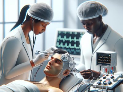
An Electroencephalogram, more commonly known as an EEG, records brain activity. It operates by placing electrodes on your scalp to capture electrical signals produced by your brain cells or neurons. These recorded waves offer valuable insights for neurologists in diagnosing conditions such as epilepsy, sleep disorders or even determining if a person is in a coma.
Take Alzheimer’s disease for instance; one study published in the Journal of Neurology reported that specific patterns found within these waveforms could indicate early signs of this debilitating condition.
What is an ECG?
On the other hand, we have Electrocardiograms—ECGs—that primarily focus on heart health instead of neurological aspects. They measure electric impulses generated when cardiac muscles contract to pump blood throughout your body.
For example, irregularities detected through an ECG can reveal whether you’ve experienced a heart attack previously—even if it was unbeknownst to you—or perhaps signal potential risks related with arrhythmia (irregular heartbeat). An article from The American Heart Association highlighted how crucial this diagnostic tool proves especially during emergency scenarios where quick decisions are paramount.
Key Differences between EEG and ECG
This section dives deeper into the contrasts that set apart an Electroencephalogram (EEG) from an Electrocardiogram (ECG), primarily focusing on their purpose, function, and data collection techniques.
Difference in Purpose and Function
An EEG’s primary objective revolves around examining brain activity. It maps out neural action by tracing electrical signals transmitted across neurons. From diagnosing epilepsy to detecting early signs of Alzheimer’s disease or sleep disorders – it plays a pivotal role.
On the flip side, an ECG maintains its focus firmly on heart health. Its main function lies in charting electric impulses produced during cardiac muscle contractions. The information garnered helps uncover traces of past heart attacks or risks related to irregular heartbeat patterns known as arrhythmias – indispensable insights for life-saving decisions in emergency situations.
Difference in Data Collection Techniques
The technique applied when collecting data via EEG involves placing electrodes directly onto your scalp – essentially mapping out electrical activity straight from your brain’s source with high temporal resolution but limited spatial accuracy due to signal distortion caused by intervening tissues like skull bone and skin layers.
In contrast, recording through ECG sees electrodes attached strategically around one’s chest area aiming at capturing every single heartbeat detail accurately while offering real-time results though restricted by lower temporal precision compared against direct intracardiac recordings.
How EEG Works
Delving into the workings of an Electroencephalogram (EEG), it’s essential to grasp how brain waves function and understand the procedure, interpretation of this diagnostic tool. This in-depth exploration can give you a better understanding about your body’s internal electrical systems.
Understanding Brain Waves
Brainwaves represent communication among billions of neurons within your brain. These patterns provide valuable insights for clinicians when analyzing an EEG test. Four primary types exist: Delta, Theta, Alpha, and Beta waves — each playing distinct roles in different mental states.
Delta waves primarily appear during deep sleep or while under anesthesia; they’re typically observed at frequencies below 4 Hz.
Theta wave activity occurs between 4-7 Hz frequency range often associated with creativity and intuitive thinking but also present during light sleep or relaxation periods.
Alpha waves are noticeable predominantly during restful moments yet alert intervals running around 8-13 Hz on average – think times where you’re peacefully reading a book.
Beta waves signify active thought processes generally falling between14–30Hz spectrum – these would be prevalent while problem-solving or making decisions.
Comprehending these signals is crucial as anomalies could indicate various neurological conditions like epilepsy or Alzheimer’s disease.
EEG Procedure and Interpretation
An efficient method for capturing neuronal activities from your scalp— that’s what makes up an electroencephalography procedure! It starts by positioning small metal discs known as electrodes across certain areas atop one’s head. A conductive gel aids in maintaining good contact allowing accurate data collection without any invasive measures needed!
Next comes recording session lasting approximately half-hour long wherein patient must stay relaxed throughout ensuring no artificial interference disrupts natural neural oscillations being tracked down meticulously by device hooked onto said electrodes feeding every bit information straight through computer system designed specifically analysing such complex electrophysiological data sets giving rise much more than just squiggly lines seen printout post examination rather comprehensive overview entire cerebral activity!
Interpreting an EEG report involves identifying patterns within the collected data. Clinicians look for abnormalities such as slow or fast brain waves, spikes in activity indicative of potential seizures, and other irregular waveforms that could suggest neurological issues.
Remember this – every individual’s EEG may vary due to factors like age, sleep status during testing, and even certain medications can influence these results.
How ECG Works
Following a detailed understanding of EEG, it’s time to shift our focus towards the workings of an Electrocardiogram (ECG). Like its counterpart, the process behind this diagnostic tool involves recording electrical activity. But, in contrast with EEGs that concentrate on brain function, an ECG is pivotal for monitoring heart health.
The Role of the Heart’s Electrical Activity
Your heart isn’t just about blood and beats; it also has a fascinating electric side. At its core lies an intricate network responsible for generating impulses at regular intervals – think of them as small shocks ensuring your heart keeps rhythmically contracting and relaxing. This continuous cycle enables vital circulation around your body: delivering oxygen-rich blood from lungs to tissues while bringing deoxygenated one back for re-oxygenation.
The central pacemaker or sinoatrial node initiates each heartbeat by firing off these electric signals which then traverse through atrial muscles before reaching another critical point –-the ventricular conductive system– causing contractions there too! And voila! You’ve got yourself a perfectly functioning pump working round-the-clock without rest!
In cases where anomalies occur in this finely tuned orchestra—for instance if parts beat out-of-sync—it can lead to potentially life-threatening conditions such as arrhythmias or cardiac arrests. That’s exactly when you’d need an electrocardiogram (ECG) test—a crucial tool diagnosing irregularities within this precise conduction pathway.
ECG Procedure and Interpretation
Having understood what makes up hearts’ electrical world let’s investigate into how medical professionals harness technology like ECG monitors revealing abnormalities lurking beneath surface rhythms we typically associate with normal “heartbeats”.
During procedure doctors strategically place 10 electrodes over patient’s chest arms legs capturing various aspects associated with impulse propagation pathways across different regions myocardium—each electrode records distinct but complementary data reflecting comprehensive panorama inherent cardiovascular dynamics present moment recorded reading.
Data obtained from these electrodes are then translated into a graph-like representation, often referred to as ECG waveforms. Each part of the waveform corresponds to different stages in the heart’s electrical cycle—P waves for atrial depolarization, QRS complex representing ventricular depolarization and T waves signifying ventricular repolarization.
Applications of EEG and ECG in Medical Practice
As we dive deeper into the applications of both Electroencephalogram (EEG) and Electrocardiogram (ECG), it’s crucial to understand their distinctive uses within medical practice.
Use of EEG in Diagnosing Neurological Disorders
An essential tool for neurologists, an EEG offers insights into brain activity that can’t be gleaned from other diagnostic procedures. Epilepsy, a neurological disorder characterized by recurrent seizures, serves as one prime example where EEGs play an instrumental role. During seizure episodes, there’s increased neuronal electrical discharge causing unusual patterns on the recorded data.
The detection isn’t limited to epilepsy; mental health conditions like depression also present altered neural oscillations visible through this procedure. For instance, Alpha waves linked with calmness might show less prevalence compared to Beta waves associated with active thinking or anxiety—indicating potential depressive states.
Sleep disorders too fall under the purview of diagnosis via electroencephalography—the process behind capturing these readings using an EEG machine—with sleep apnea being commonly detected due its effects on normal sleep cycle stages reflected upon corresponding wave types: Theta during light slumber and Delta while deeply asleep.
Use Of ECG In Cardiovascular Health Assessment
In terms cardiovascular diagnostics lies another arena where monitoring electric impulses is critical—that’s precisely what an ECG does—it gauges cardiac performance through mapping heartbeats onto interpretable graphs known as cardiographs or simply ‘tracings’.
Arrhythmias—irregularities disrupting rhythmical contractions—are often diagnosed via carefully examining these tracings since every component represents specific phases: P-wave denotes atrial depolarization before heartbeat initiation whereas QRS complex signifies ventricular contraction marking actual heartbeat onset itself.
Yet arrhythmia isn’t sole concern here—a history involving myocardial infarction better known as heart attack leaves distinct scars detectable even long after the event via particular ECG wave patterns—Q-waves. This detection becomes vital in proactive healthcare management, particularly amongst patients with higher risk profiles.
Also, these graphs also provide evidence of ongoing heart conditions like ischemia or angina—a condition causing chest pain due to insufficient blood supply to cardiac muscles often resulting from coronary artery blockages detectable through ST-segment deviations on an ECG tracing.
In essence, while EEG and ECG share a common foundation—that is recording electrical activity—they cater different aspects within medical field: neurological versus cardiovascular respectively; each having their own set diagnostic applications which are indispensable for effective patient care.
Pros and Cons of EEG and ECG
After diving deep into the primary functions, techniques, and applications of both Electroencephalogram (EEG) and Electrocardiogram (ECG), let’s shift our focus to understand their benefits along with limitations. By acknowledging these aspects, you can comprehend why medical professionals choose one over the other based on patient needs.
Benefits of EEG
In clinical practice, an EEG provides numerous advantages. Its high temporal resolution enables it to capture brain activity in milliseconds – a critical aspect when diagnosing conditions like epilepsy where seizure events are short-lived yet impactful. Besides this remarkable speed factor, its non-invasive nature adds another advantage as patients aren’t exposed to any risks related from invasive procedures such as lumbar punctures or biopsies.
Also, being relatively inexpensive compared to imaging technologies like MRI or CT scans makes it accessible for various healthcare settings – be that clinics operating under tight budgets or countries with limited resources.
Benefits of ECG
An essential player in cardiovascular health assessment is none other than ECGs due largely to their simplicity paired up with profound implications they hold for heart disease detection including arrhythmias or past myocardial infarctions which can save lives if detected early enough before causing severe damage towards heart muscles.
Apart from detecting potential threats through specific wave patterns seen during examination phase ,their fast response times make them suitable choice especially emergency situations while offering real-time data helps doctors decide course action immediately without losing crucial time waiting results come back laboratory tests.
Limitations of EEG
While providing key insights about neurological functioning via electrical signals captured by scalp-placed electrodes —an approach commonly referred as passive electrode placement—this technique has certain drawbacks too mainly linked spatial accuracy since these surface level recordings often get blurred due diffused neural activities happening deeper inside brain making harder discern exact origin abnormal signal propagation within cerebral cortex region also known ‘source localization’ problem neuroimaging field.
Besides, external factors like patient movements or eye blinks can distort the EEG signal and produce false readings. Also, interpreting these signals requires specialized training as normal brain activity varies across individuals based on age, sleep state etc., making it a subjective interpretation susceptible to errors.
Limitations of ECG
Even though its invaluable contributions towards cardiac care arena ,there are certain limitations associated with ECG testing process including potential misinterpretation waveforms by medical practitioners due complex nature heart’s electrical system which might lead incorrect diagnosis sometimes .
Also,the limited temporal resolution compared direct intracardiac recordings also presents drawback context detailed heartbeat analysis . Also,body size variations among patients influence accuracy electrode placements hence altering quality data obtained from procedure besides possible discomfort caused during application electrodes chest arms legs especially cases where hair needs be shaved off ensure proper contact skin surface .
Conclusion
You’ve now navigated the differences between EEG and ECG, two critical diagnostic tools in healthcare. It’s clear that while both record electrical activity, they have unique applications—EEG for neurological issues like epilepsy or sleep disorders, and ECG for diagnosing heart-related problems such as arrhythmias. Remember an EEG leverages electrodes on your scalp to monitor brain activity while an ECG captures heartbeat details using strategically placed chest electrodes.
Their pros and cons further differentiate them with EEG being cost-effective yet potentially affected by external factors; whereas an ECG provides rapid results but may present discomfort during electrode placement. Understanding these key distinctions can empower you not only when dealing with health concerns but also enhancing your appreciation of our body’s complex systems. So next time someone confuses their names due to similarity remember – one scans the rhythm of thought patterns (EEG) another maps out the beats of life itself (ECG).
- Xbox Versus PlayStation: A Comparative Analysis - November 15, 2025
- GDP Versus GNP: Understanding the Key Differences - November 14, 2025
- Which Is Best: CV or Resume? Understanding the Differences - November 14, 2025


