Exploring medical imaging technologies can sometimes feel overwhelming. With multiple available methods designed to assess similar health concerns, understanding the nuanced differences becomes essential. Among echocardiographic techniques, Transthoracic Echocardiography (TTE) and Transesophageal Echocardiography (TEE) stand out as remarkable tools designed for unique clinical applications. This discussion delves into their specific functionalities, contrasts, and the considerations influencing their selection.
Overview of TTE and TEE
Definition and Purpose of TTE
TTE, standing for Transthoracic Echocardiography, represents a non-invasive diagnostic tool used to visualize the heart’s structure and assess its function. Utilizing ultrasonic waves transmitted via an externally placed transducer, TTE provides detailed images that assist clinicians in identifying cardiac abnormalities efficiently and safely.
Definition and Purpose of TEE
Transesophageal Echocardiography, abbreviated as TEE, involves the insertion of a specialized transducer into the esophagus. Situated closer to the heart, TEE enables acquisition of precise and highly detailed images, especially useful when standard transthoracic views are insufficient or obstructed.
Differences Between TTE and TEE
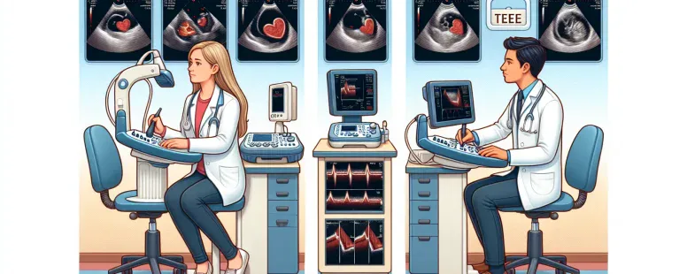
Imaging Techniques and Capabilities
TTE relies on surface-level imaging, with the ultrasound waves penetrating through the chest wall to capture cardiac views. Conversely, TEE positions the transducer within the esophagus, resulting in superior resolution and access to regions obscured in TTE imaging.
Applications and Indications
TTE serves as a first-line echocardiographic evaluation method for common cardiac conditions, whereas TEE is preferred for detailed assessments of intricate structures like valves and atria or when TTE imagery is inconclusive.
Implementation Procedures and Patient Experience
TTE Procedure Overview
The TTE procedure entails a non-invasive process where a transducer probe is gently moved across the chest to obtain various cardiac views. It is generally quick, causes minimal discomfort, and is highly accessible.
TEE Procedure Overview
In contrast, the TEE procedure necessitates sedation, followed by the insertion of the transducer into the esophagus. Though the images produced are unparalleled in clarity, the procedure requires more preparation and post-procedure observation due to the sedation involved.
Advantages and Limitations
TTE: Benefits and Drawbacks
TTE is celebrated for its ease of implementation, non-invasive nature, and widespread availability. But, its imaging capability may be limited in patients with obesity or conditions restricting chest wall imaging.
TEE: Benefits and Drawbacks
TEE compensates for TTE’s limitations with its superior imaging of hard-to-reach cardiac areas, critical for surgeries or detecting complex pathologies. Nevertheless, it is more invasive and requires expert handling.
Factors Influencing Choice Between TTE and TEE
Clinical Case Studies Highlighting TTE and TEE Use
Specific case studies underscore the importance of selecting the appropriate technique. For example, TTE is often sufficient for initial assessments, but TEE becomes indispensable in guiding specific surgical interventions or defect identifications when clarity is paramount.
Published: November 13, 2025 at 4:32 pm by Ellie B, Site owner & Publisher
To provide the best experiences, we use technologies like cookies to store and/or access device information. Consenting to these technologies will allow us to process data such as browsing behaviour or unique IDs on this site. Not consenting or withdrawing consent, may adversely affect certain features and functions.
Functional
Always active
The technical storage or access is strictly necessary for the legitimate purpose of enabling the use of a specific service explicitly requested by the subscriber or user, or for the sole purpose of carrying out the transmission of a communication over an electronic communications network.
Preferences
The technical storage or access is necessary for the legitimate purpose of storing preferences that are not requested by the subscriber or user.
Statistics
The technical storage or access that is used exclusively for statistical purposes.The technical storage or access that is used exclusively for anonymous statistical purposes. Without a subpoena, voluntary compliance on the part of your Internet Service Provider, or additional records from a third party, information stored or retrieved for this purpose alone cannot usually be used to identify you.
Marketing
The technical storage or access is required to create user profiles to send advertising, or to track the user on a website or across several websites for similar marketing purposes.