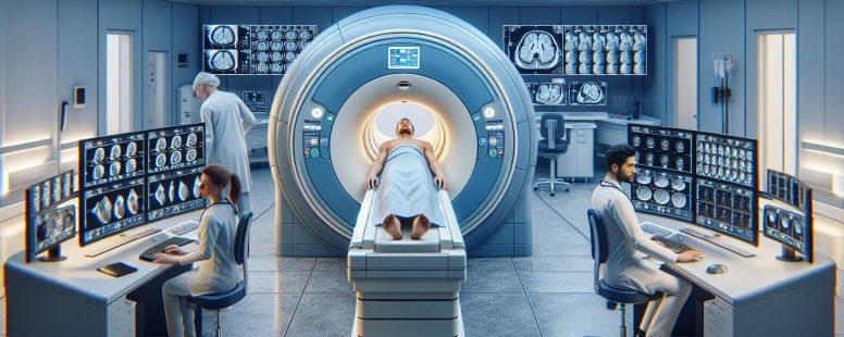
Ever found yourself puzzled by the medical jargon tossed around in a doctor’s office or during a casual conversation about health? You’re not alone. Two terms that often cause confusion are MRI and fMRI. These acronyms represent powerful imaging technologies, but what sets them apart?
Understanding MRI and fMRI
Building on the understanding of medical imaging technologies, let’s investigate into Magnetic Resonance Imaging (MRI) and Functional Magnetic Resonance Imaging (fMRI). These two methods, though similar in name, have distinct functionalities.
What is MRI?
Magnetic Resonance Imaging or MRI represents a non-invasive technique that doctors employ to inspect the insides of your body. It utilizes a large magnet and radio waves to generate detailed images of organs and structures within your body.
Let’s take an example: If you’re experiencing persistent headaches, physicians may use an MRI scan to check for any abnormalities in brain tissues like tumors or inflammation—providing accurate information about physical health conditions.
What is fMRI?
Functional Magnetic Resonance Imaging—shortened as fMRI—is another type but it dives deeper than its counterpart by analyzing blood flow changes related directly with neural activities inside the brain. This means instead of only producing static images, it provides dynamic ones which indicate how different parts interact while performing specific tasks or during rest periods.
The Technical Difference Between MRI and fMRI
Digging deeper into the technicalities, you’ll find significant differences between Magnetic Resonance Imaging (MRI) and Functional Magnetic Resonance Imaging (fMRI). Let’s explore these under two key areas: magnetization process in both procedures and image acquisition techniques.
Magnetization in MRI and fMRI
In an ordinary MRI procedure, a strong magnetic field aligns hydrogen atoms within your body. These aligned atoms then absorb radio waves that trigger their rotation. When the radio wave source is turned off, these spinning atoms return to their original position by emitting energy captured as signals which create detailed images of your body tissues.
Contrarily, in an fMRI scan there isn’t just focus on water molecules or atomic alignment but it also detects changes associated with blood flow. This technique leverages Blood Oxygen Level Dependent (BOLD) contrast that associates brain activity levels with alterations in oxygenated versus deoxygenated blood ratio.
Image Acquisition in MRI and fMRI
For traditional MRIs, once magnetized by powerful fields those emitted energies from rotating hydrogen ions are measured using receiver coils strategically placed around target area of examination like head for neurological scans or abdomen for gastrointestinal investigations etc., later translated into high resolution static images portraying anatomical details vividly.
Functional vs Structural Imaging
Exploring the distinctions between MRI and fMRI further, it’s vital to understand how each type of imaging contributes uniquely to medical diagnostics. This section focuses on illuminating this contrast by detailing how MRI provides structural imaging while fMRI captures functional data.
How MRI Provides Structural Imaging
An essential aspect of Magnetic Resonance Imaging (MRI) lies in its ability for structural visualization. By creating detailed pictures of organs and tissues within your body, MRIs help identify any potential abnormalities or issues with precision.
Let’s jump into an example: consider a patient experiencing chronic headaches without apparent cause; An MRI can be instrumental here—its high-resolution images assist doctors in identifying possible sources such as tumors or inflammation that may not have been detected otherwise.
Unlike other forms of diagnostic tools like X-rays or CT scans which only provide two-dimensional views, an MRI goes beyond – delivering three-dimensional imagery from various angles so providing unparalleled anatomical detail.
How fMRI Captures Functional Imaging
On the flip side is Functional Magnetic Resonance Imagining (fMRI). Rather than just showcasing static images like traditional MRIs do, this technology delves deeper—it monitors dynamic changes occurring inside your brain over time due to different activities you engage in or even when at rest!
A fascinating feature about these scans revolves around their capability called Blood Oxygen Level Dependent (BOLD) contrast – they’re able to link variations between oxygenated and deoxygenated blood with specific levels of brain activity—an invaluable asset indeed for neurologists studying cognitive function disorders including Alzheimer’s disease and schizophrenia among others.
Uses and Applications of MRI and fMRI
Diving deeper into the practical aspects, let’s look at how these imaging techniques are employed in real-world scenarios. The uses of both MRI and fMRI stretch far beyond just capturing images; they play crucial roles in diagnosis, research, treatment planning, and monitoring.
Medical Applications of MRI
Considered a mainstay for many medical diagnoses due to its high-resolution imagery capabilities, an MRI proves instrumental across various fields. In neurology it helps detect conditions like multiple sclerosis by revealing plaques or scarring on brain tissue. It aids orthopedic assessments as well – illustrating torn ligaments or cartilage within joints such as knees or shoulders.
Radiologists often employ MRIs when examining organs – kidneys harboring cysts for instance get identified efficiently through this technique. When dealing with cardiovascular ailments too, cardiologists find MRIs useful because it depicts heart structures without needing invasive procedures.
Finally yet importantly lies oncology: using contrast agents during an MRI allows detection of tumors accurately while avoiding radiation exposure inherent in other modalities like CT scans.
Research and Clinical Uses of fMRI
While regular MRIs excel at structural visualization,fMRIs shine where dynamic functionality is concerned especially about neurological studies.
Researchers use fMRis frequently to observe different areas’ responses inside the brain when performing specific tasks.This understanding paves ways towards studying cognitive disorders including autism spectrum disorder(ASD), attention deficit hyperactivity disorder (ADHD) among others.It provides invaluable insights concerning Alzheimer’s disease,schizophrenia since anomalies can be seen related to oxygen levels coupled with activities conducted by patients simultaneously.
Also,in clinical settings doctors use FMRI before surgeries that involve delicate parts around our brains.For example,it assists surgeons identify critical regions tied up with speech,movement control so unnecessary damages could be prevented.So,this modality remains vital not only mapping out our minds but also ensuring patient safety throughout surgical interventions.
Safety and Side Effects
After understanding the core differences between MRI and fMRI, it’s crucial to discuss their safety aspects. Each procedure comes with a unique set of considerations about patient safety.
Safety Concerns of MRI
One significant concern associated with MRIs relates to magnetic fields’ strength used during scanning. High-powered magnets can cause metal objects within or on patients—such as pacemakers, cochlear implants, or certain types of vascular clips—to move violently or malfunction. Hence, all metallic items must be removed before an MRI scan.
Exposure to loud noises during scans is another aspect requiring attention in MRIs. The banging sound produced by gradient coils inside the scanner may result in temporary hearing loss if appropriate ear protection isn’t provided.
In rare instances (less than 1% according to RadiologyInfo.org), patients might experience allergic reactions from contrast agents administered for enhancing images captured by the machine.
Side Effects of fMRI
While generally considered safe because they don’t use ionizing radiation like CT scans do — no long-term health risks have been documented concerning repeated exposure — functional MRIs aren’t without potential side effects either.
The most common issue that could occur is claustrophobia due its enclosed structure where some people report feeling anxious when confined in tight spaces such as an fMRI tube-like device which results into panic attacks occasionally hence making them unable complete imaging process smoothly .
Also , similar traditional MR Imaging technology there’s also risk related high-strength magnet causing problems those who wear medical devices metallic components along concerns about noise levels too but compared less severe since typically shorter duration uses lower field strengths overall increasing patient comfort significantly .
Conclusion
You’ve now navigated the complex world of MRI and fMRI, understanding their unique roles in modern medicine. You know that while both are advanced imaging technologies, they serve distinct purposes. The former gives us a detailed static picture of body structures to spot abnormalities like tumors or inflammation; the latter brings brain activities into focus by tracking changes in blood flow linked with neural activity levels.
Diving deeper, you grasped how technical differences underpin these functionalities – from magnetization processes to image acquisition techniques. Also, you saw firsthand how each method’s practical applications extend beyond mere theory – impacting fields such as neurology and oncology for MRIs or neurological research and surgery planning for fMRIs.
Finally, being aware of safety considerations ensures your informed engagement during medical procedures involving these tools. So whether it’s spotting potential health risks through an MRI scan or uncovering intricate workings of our brains via an fMRI study – remember, this knowledge empowers your healthcare journey!
To provide the best experiences, we use technologies like cookies to store and/or access device information. Consenting to these technologies will allow us to process data such as browsing behaviour or unique IDs on this site. Not consenting or withdrawing consent, may adversely affect certain features and functions.
Functional
Always active
The technical storage or access is strictly necessary for the legitimate purpose of enabling the use of a specific service explicitly requested by the subscriber or user, or for the sole purpose of carrying out the transmission of a communication over an electronic communications network.
Preferences
The technical storage or access is necessary for the legitimate purpose of storing preferences that are not requested by the subscriber or user.
Statistics
The technical storage or access that is used exclusively for statistical purposes.The technical storage or access that is used exclusively for anonymous statistical purposes. Without a subpoena, voluntary compliance on the part of your Internet Service Provider, or additional records from a third party, information stored or retrieved for this purpose alone cannot usually be used to identify you.
Marketing
The technical storage or access is required to create user profiles to send advertising, or to track the user on a website or across several websites for similar marketing purposes.